Module 1: The Nervous System
Module Introduction
The nervous system is the main communication and control system of the body. The nervous system is responsible for the production of movement and emotive behaviour as well as the experience of sensory events (both physical and emotional).
In this module, we discuss the two major divisions of the nervous system and introduce some of the structural elements of the nervous system involved in the control and co-ordination of body activities.
Module Structure
This module contains two sections:
Section 1: Organization of the Nervous System
Section 2: Neurotransmission
Section 1 – Organization of the Nervous System
Learning Objectives
After reading this section, you should be able to:
- State the general functions of the nervous system.
- Identify the components of the central nervous system and the peripheral nervous system.
- Describe the main functions of the components of the central and peripheral nervous systems.
- State the function of the somatic and autonomic nervous systems.
- State the function of the structural components of a neuron.
- Describe the function of the three types of nerves.
- Describe the function of the varioustypes of neuroglia.
General Functions of the Nervous System
The nervous system is the main communication and control system of the body. It is responsible for the production of movement, sensation, reasoning, memory and emotion. The primary functions of the nervous system include communication, integration and control of motor (movement), sensory and behavioural activities. The nervous system co-ordinates the body’s conscious and unconscious responses to stimuli.
The overall function of the nervous system is to:
-
-
- receive information from the environment and the body
- analyze and process this information
- initiate a response or plan of action by transmitting electrical impulses* through interconnected nerve cells
-
Many of the functions of the nervous system occur automatically in response to various stimuli.
Voluntary actions are initiated by higher, conscious areas of the brain.
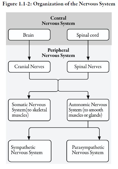
Organization of the Nervous System
The components of the nervous system can be grouped according to structure or function.
Anatomically, the nervous system is subdivided into two major divisions: (see Figure 1.1-1)
- central nervous system*
- peripheral nervous system*
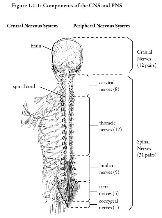
1) Central nervous system (CNS)
The CNS consists of the spinal cord and the brain. The brain receives and processes information derived from the environment and the rest of the body, makes decisions, and sends “commands” for action. The brain relies on the peripheral nervous system (PNS) to receive its information. The spinal cord serves as a relay station between the brain and the periphery.
2 Peripheral nervous system (PNS)
The PNS refers to nerves and nerve tissue that fan out from the brain and spinal cord:
-
-
- cranial nerves – 12 pairs of cranial nerves emerge directly from the brain
- spinal nerves – 31 pairs of spinal nerves (cervical, thoracic, lumbar, sacral, and coccygeal) are connected to the spinal cord
- ganglia* – clusters of nerve cell bodies that are associated with the spinal nerves
-
The PNS links the brain and spinal cord to peripheral tissues such as skeletal muscles and viscera (internal organs).
The PNS transmits information derived from body organs and the environment to the brain, either directly or via the spinal cord, and from the brain to the muscles and organs. In other words, the peripheral nervous system relays information to the CNS and then transmits information from the CNS back to the muscles and organs.
The peripheral nervous system structures can be further subdivided into two main divisions:
-
-
- somatic* nervous system
- autonomic* nervous system
-
In the somatic (body) nervous system, there are both sensory (afferent*) nerves and motor (efferent*) nerves. Somatic sensory nerves conduct impulses from the skin, muscles and joints to the CNS to enable sensation of body parts. Somatic motor nerves conduct impulses from the CNS to the skeletal muscles of the trunk and limbs to permit voluntary movement.
The autonomic (self-regulating) nervous system refers to the part of the nervous system involved mainly in regulation of visceral (internal) organs and smooth muscles, which play an important role in preparing the body to respond quickly to environmental stimuli. The autonomic nervous system regulates automatic or involuntary processes such as heart rate, pupillary dilation, gastrointestinal tract movement and urinary bladder function. The autonomic nervous system is composed of two distinct subsystems that are highly interdependent and counterbalance each other:
-
-
- sympathetic nervous system* – activates organ activity to respond quickly to threatening stimuli, prepares the body for “fight or flight”
- parasympathetic nervous system* – returns organ activity to resting state after the threatening situation is resolved (restoring and maintaining homeostasis)
-
For example, if you were in a frightening situation, the sympathetic nervous system would cause your heart rate to increase. After the threatening situation has passed, the parasympathetic nervous system would slow your heart rate.
We discuss the central nervous system in more detail in Module 2, and the peripheral nervous system in Module 3.
Neurons
The neuron*, or nerve cell, is the basic unit of the nervous system. Each neuron is a microscopic information processing system that receives and transmits messages to and from other cells.
Neurons have a nucleus, a membrane, and a variety of structures within the cell body, just as other cells do. However, neurons, unlike many other cells, do not possess the ability to regenerate under normal circumstances. This explains why damage to nerve structures may lead to permanent sequelae. Also, neurons do not look like other cells because they have dendrites* and axons* extending from the body of the cell.
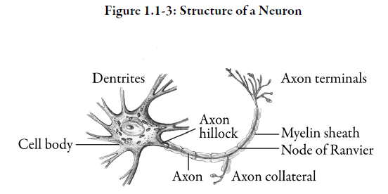
Dendrites are located at the receptive end of the neuron and conduct impulses toward the body of the nerve cell. Depending on the type of neuron, it may have hundreds of branching dendrites. Within the cell body, the signals received are integrated to produce a single output to the axon.
Axons are nerve fibres that conduct impulses away from cell bodies to the target cells or tissues. Therefore, neurons are described as having a bipolar structure. Signals are received at one end of the cell and output is transmitted from the other end.
Each neuron has only one axon. However, depending on the location of the neuron, its axon may be very short (< 1 mm) or very long (e.g., extending from the lumbar spine to the toe). The area where the axon emerges from the cell body is called the axon hillock*. This part of the neuron has the greatest density of voltage-dependent sodium channels and is the most easily excited part of the neuron.
Most axons are coated with a fatty, insulating layer called a myelin sheath*. The presence of myelin increases the speed of transmission of electrical impulses along the axon, and also helps prevent loss of electrical charge. The myelin sheath is punctuated by nodes of Ranvier*. These nodes lack myelin but contain a high density of voltage-gated ion channels.
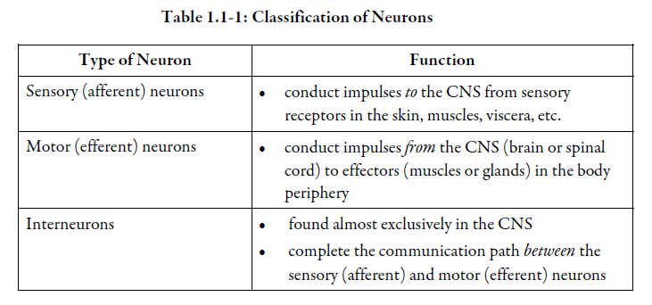
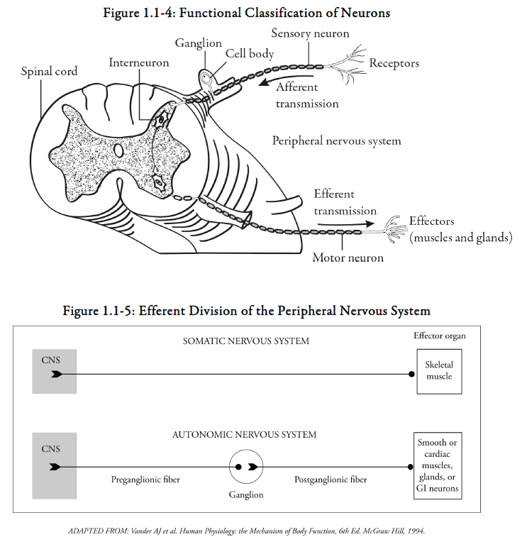
Neurons are classified according to the direction in which they transmit impulses. See Table 1.1-1 and Figure 1.1-4.
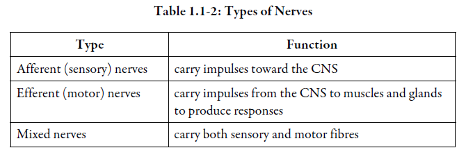
In the somatic nervous system, there is only one neuron between the CNS and the effector organ (e.g., skeletal muscle). In the autonomic nervous system, there are two neurons between the CNS and effector organ (e.g., viscera). The neurons of the autonomic nervous system meet in an outlying ganglion before they reach their target organ. Preganglionic fibres* are axons that are located before the ganglia. Postganglionic fibres* are located after the ganglia. See Figure 1.1-5.
Nerves
Nerves are bundles of axons that follow a common pathway. To appreciate the difference between a nerve and a neuron the comparison is often made to wires versus cables. A neuron may be considered an individual wire that conducts an electrical impulse, whereas a nerve is like a cable carrying hundreds or thousands of wires. Nerves can be seen by the naked eye; however, neurons can only be visualized under the microscope.
Nerves, like neurons, are classified according to the direction in which they transmit impulses. See Table 1.1-2.
-
-
- afferent, or sensory nerves – carry impulses toward the CNS
- efferent or motor nerves – carry impulses from the CNS to muscles and glands to produce responses
- mixed nerves – carry both sensory and motor fibres
-
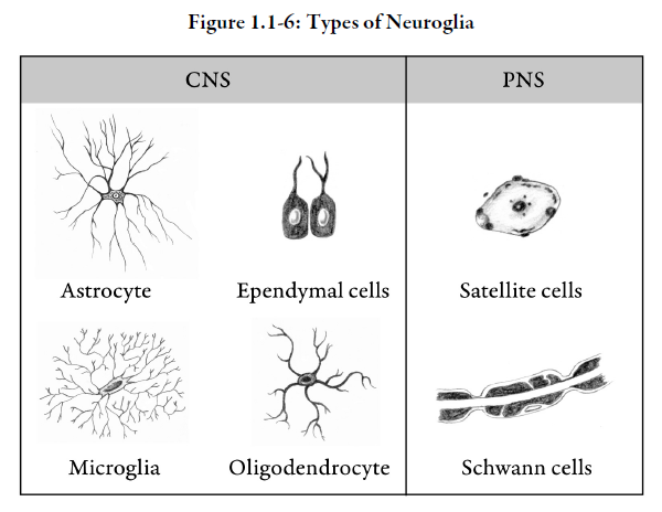
Neuroglia
In addition to neurons, nervous tissue is also composed of neuroglia*. Neuroglia is sometimes referred to as “nerve glue”. There are two significant differences between neurons and neuroglia. Unlike neurons, neuroglia do not transmit nerve impulses. Neuroglia can replicate; most neurons cannot.
Cells of the neuroglia are commonly referred to as glial cells. There are several types of glial cells. See Table 1.1-3 and Figure 1.1-6.
Summary — Section 1: Organization of the Nervous System
The nervous system is the main communication and control system of the body. Anatomically, it is subdivided into the central nervous system (CNS) and the peripheral nervous system (PNS). The spinal cord and the brain make up the central nervous system. The peripheral nervous system is comprised of the cranial nerves, spinal nerves, ganglia and peripheral receptors. They link the brain and spinal cord to peripheral tissues.
The peripheral nervous system is further subdivided into the somatic (voluntary) nervous system and the autonomic (self-regulating) nervous system. In the somatic nervous system, sensory (afferent) nerves transmit information to the CNS from skin, muscles and joints. Motor (efferent) nerves conduct impulses from the CNS to skeletal muscles of the trunk and limbs to permit voluntary movement.
The autonomic nervous system is involved in the self-regulation of visceral function. It is composed of the sympathetic nervous system and the parasympathetic nervous system, which counterbalance each other. The autonomic nervous system regulates a number of visceral organs and glands including heart rate, pupillary dilation, gastrointestinal tract movement and urinary bladder function.
The neuron is the functional unit of the nervous system. Each neuron contains a cell body, several dendrites and one axon.
Nerves are bundles of axons. Nerves are classified as:
-
-
- afferent or sensory nerves
- efferent or motor nerves
- mixed nerves
-
Neuroglia are cells found in nerve tissue. They do not transmit nerve impulses. In the CNS, neuroglia include astrocytes, microglia, oligodendrocytes and ependymal cells. In the PNS, Schwann cells and satellite cells are types of neuroglia.
Progress Check — Section 1: Organization of the Nervous System
1.
List the three main functions of the nervous system.
1 _____________________________
2 _____________________________
3 _____________________________
2.
The CNS is composed of:
a. the brain and spinal cord
b. the brain, spinal cord and skeletal muscles
c. the somatic and autonomic nervous systems
d. the cell body, axons and dendrites
3.
The brain receives its information from the _________________________ nervous system.
4.
Nerves that conduct impulses from the skin, muscles, and joints to the CNS are called _________________________ nerves.
5.
Nerves that permit conscious control of skeletal muscle are called ______________________ nerves.
6.
The subdivision of the nervous system involved with self-regulation of visceral organs is called the _________________________.
7.
The parasympathetic nervous system and the sympathetic nervous system are the two subdivisions of the ___________________________ nervous system.
8.
Neurons differ from other body cells in that each neuron has:
a. a nucleus in the cell body
b. a cell membrane
c. branches that extend away from the cell body
d. a variety of structures within the cell
9.
Nerve fibres that conduct impulses away from the cell body of a neuron are called _________________________.
10.
How many axons does a neuron typically have?
___
11.
Nerves are defined as bundles of axons that follow a common pathway.
a. true
b. false
12.
What are the three types of nerves?
1) _______________________
2) _______________________
3) _______________________
13.
Which glial cells act as phagocytes?
a. astrocytes
b. microglia
c. oligodendrocytes
14.
How do neurons and neuroglia differ?
_________________________
_________________________
Progress Check Answers — Section 1: Organization of the Nervous System
1.
1) receive information from the environment and the body
2) analyze and process this information
3) initiate a response
2.
a. the brain and spinal cord
3.
peripheral
4.
sensory (afferent)
5.
motor (efferent)
6.
autonomic nervous system
7.
autonomic
8.
c. branches that extend away from the cell body
9.
axons
10.
1
11.
a. true
12.
1) afferent (sensory)
2) efferent (motor)
3) mixed (afferent and efferent)
13.
b) microglia
14.
Neuroglia do not transmit nerve impulses; neurons do.
Neuroglia are capable of replication; most neurons are not.
Section 2 – Neurotransmission
Learning Objectives
After reading this section, you should be able to:
- Explain how a nerve impulse is transmitted.
- Describe the role of neurotransmitters at the synapse.
- Describe the function of the common neurotransmitters.
Neurotransmission
Neurotransmission refers to the transmission of an electrical impulse along the axon of a neuron to target cells or tissues. The impulse travels along the membrane of the axon. This membrane lets in some charged particles (ions) and keeps others out. Electrical properties related to the nerve membrane can be divided in four states, as follows:
1) Resting state (polarized)
In the resting state or resting potential (i.e., the neuron is not being stimulated), the inside of the axon is negatively charged with respect to the outside and the membrane is said to be polarized. The major extracellular ion that contributes to the resting potential is sodium (Na+); the major intracellular ion is potassium (K+). Ion gradients of concentration are maintained by the action of ionic pumps that pump out Na+ and pump in K+ across the cell membrane. These different ion types and their chemical gradients between the inside and outside of the cell membrane generate a local electrical current that determines the resting potential. See Figure 1.2-1, #1.
2) Action potential (depolarization)
When a part of the axon is stimulated, the permeability of the membrane is changed, allowing sodium ions to flood into the axon. This causes a local reversal of charge and the cell is said to be depolarized. The electrical impulse, also referred to as an action potential*, has begun. See Figure 1.2-1, #2.
3) Action potential (electrochemical wave)
The action potential in one area of the axon acts as a stimulus for another action potential in the next area of the axon and the process repeats itself. This causes the action potential to move down the axon in the form of an electrochemical wave. See Figure 1.2-1, #3.

Did You Know?
The threshold for an action potential is typically about -55mV (millivolts). As sodium ions flow into the cell, the membrane potential changes from -55mV to +30mV.
The wave travels in one direction since a new action potential can only be generated in an area of the axon that is in its resting state (or polarized).
Action potentials only occur when the net movement of positive charge through the ion channels is inward. The point at which this occurs is known as the threshold. As long as the stimulus is strong enough to exceed this threshold, the action potential will occur. Stronger stimuli will not result in larger action potentials. This property of action potentials occurring maximally or not occurring at all is sometimes referred to as an all-or-none principle.
4) Repolarization
Following depolarization, potassium channels open and positively-charged potassium ions pass out of the axon. This causes the inside of the axon to become more negatively charged once again and the cell is said to be repolarized. See Figure 1.2-1, #4.
5) Hyperpolarized state
Because potassium channels close slowly, the membrane potential goes beyond (undershoots) the resting potential. The axon becomes hyperpolarized. During this phase, the nerve cell is less sensitive to further stimulation.
Opening of negatively-charged chloride ion channels by some chemical messengers lets chloride ions pass into the axon. This also causes hyperpolarization. As a result, the threshold to generate an action potential is increased and the cell is less sensitive to depolarization. See Figure 1.2-1, #4.
Finally, the sodium and potassium pump, using cellular energy in the form of ATP* (adenosine triphosphate), pumps excess sodium ions out of the cell and potassium ions back into the cell. This restores the initial concentration of sodium and potassium ions inside and outside of the nerve cell. See Figure 1.2-1, #5.
6) Refractory period
During an action potential, a second stimulus will not produce a second action potential, no matter how strong the second stimulus is. The membrane is said to be in its absolute refractory period.
After the absolute refractory period, a second action potential can be produced if the stimulus is considerably stronger than the threshold. This is the relative refractory period.
Synapse
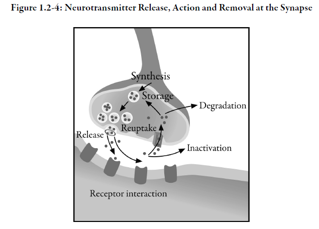
Each neuron is a separate cell. Terminals at the end of the axon link a neuron to other neurons. The axon terminals have a characteristic bead-like appearance. These are called the synaptic end bulbs. The space between the terminal on the axon of one neuron and the dendrites of another, across which the nerve impulse must travel, is known as a synapse* (or synaptic cleft). A presynaptic neuron conducts an impulse toward a synapse. An impulse is conducted away from a synapse by a postsynaptic neuron. See Figure 1.2-2.
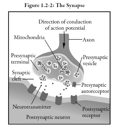
Vesicles located in terminals in presynaptic neurons are filled with neurotransmitters*, which are specialized chemical messengers that are stored and released in response to nerve stimulation.
When the action potential moving along the axon reaches the terminal of the presynaptic axon, it causes opening of voltage-gated calcium channels, which facilitates presynaptic vesicles to merge with the membrane and burst open and release neurotransmitters into the synaptic cleft. The neurotransmitter binds to receptors* on the dendrite of a receiving cell (a postsynaptic cell) and an electrochemical wave is initiated in that neuron. This process of signal passage from presynaptic to postsynaptic cells continues until the electrical impulse reaches its destination. In summary, electrical impulses are propagated from one neuron to another by the release of messenger chemicals or neurotransmitters into the synapse.
After interacting with the receptor on the receiving cell, the neurotransmitter is either destroyed by enzymes, or there is reuptake of the neurotransmitter by the presynaptic vesicles and the neurotransmitter is stored for further use.
Not every receptor contacted by neurotransmitters passes on the electrical message. Many synapses are in fact inhibitory and prevent the receiving cell from firing. A single neuron may have many excitatory and inhibitory synapses and a constant interplay between these receptor types determines whether a particular neuron fires (passes on the impulse) or not.
In addition to postsynaptic receptors, there are also presynaptic receptors on the terminal buttons of some neurons. These presynaptic receptors monitor the concentration of neurotransmitter within the synaptic cleft, providing feedback which controls the further synthesis, release and degradation of neurotransmitters. They are referred to as autoreceptors*.
This complex electrochemical process, occurring in billions of neurons, regulates the functions of the nervous system. Unfortunately, it may become dysfunctional through a number of mechanisms.
Did You Know?
The fugu fish (pufferfish) contains a toxin called tetrodotoxin that can be deadly if ingested. The toxin interrupts action potentials by blocking sodium channels. Fugu is considered a delicacy in Japan, but very few cooks know how to prepare it safely. Chefs must be specially licensed to serve fugu on their menu.
Abnormalities are known to occur when specific neurotransmitters are:
-
-
- not synthesized at all
- synthesized in insufficient amounts
- synthesized in excessive amounts
- not able to produce the desired receptor effect due to decreased numbers of receptors or receptor insensitivity
- not degraded or taken back up into the presynaptic vesicles at the normal rate
-
Also, if the amount of calcium or potassium in the synaptic region (the electrolytes* that are involved in action potential propagation and stimulation of vesicle release) changes slightly, then fewer vesicles will be activated and transmission may not occur.
When these abnormalities occur, they may result in neurological sequelae such as seizures* or abnormal movement disorders. For example, Alzheimer’s disease is associated with a deficiency of the neurotransmitter acetylcholine (ACh)* and Parkinson’s disease* is associated with a deficiency of dopamine*. Abnormalities in neurotransmitter systems also contribute to psychiatric disorders such as depression*, bipolar disorder* or other disturbances of thought processing, mood or behaviour. In this course, we focus on various neurologic disorders. Psychiatric disorders are discussed in the CE Psychiatry course.
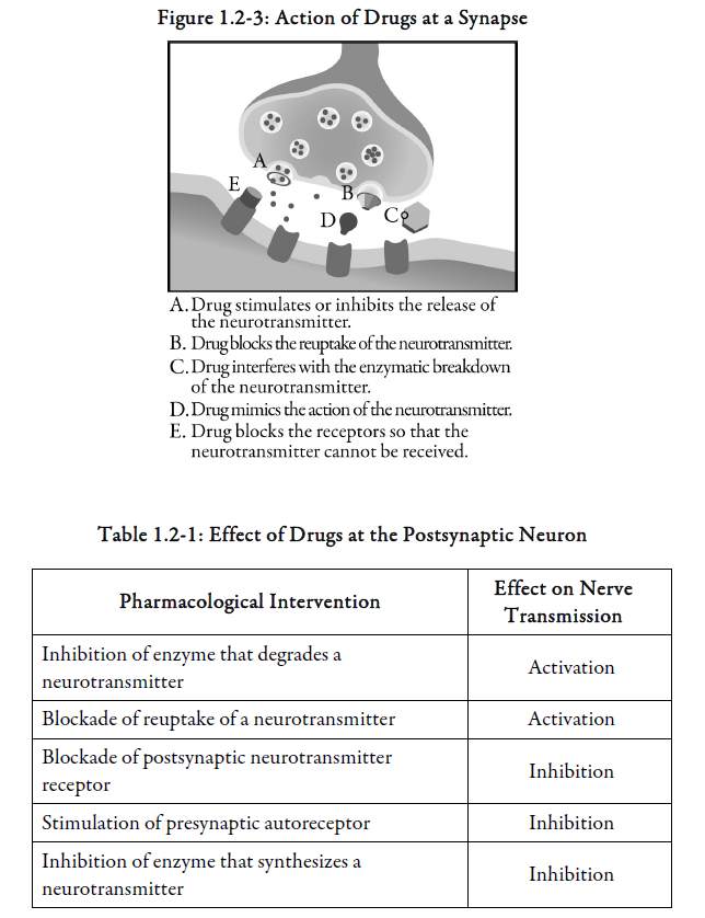
Most nervous system drugs act at the level of the synapse by modifying the activity of neurotransmitters involved in information transfer between neurons. See Figure 1.2-3 and Table 1.2-1 for the action of drugs at a synapse. Generally, only one of these actions occurs at a particular synapse.
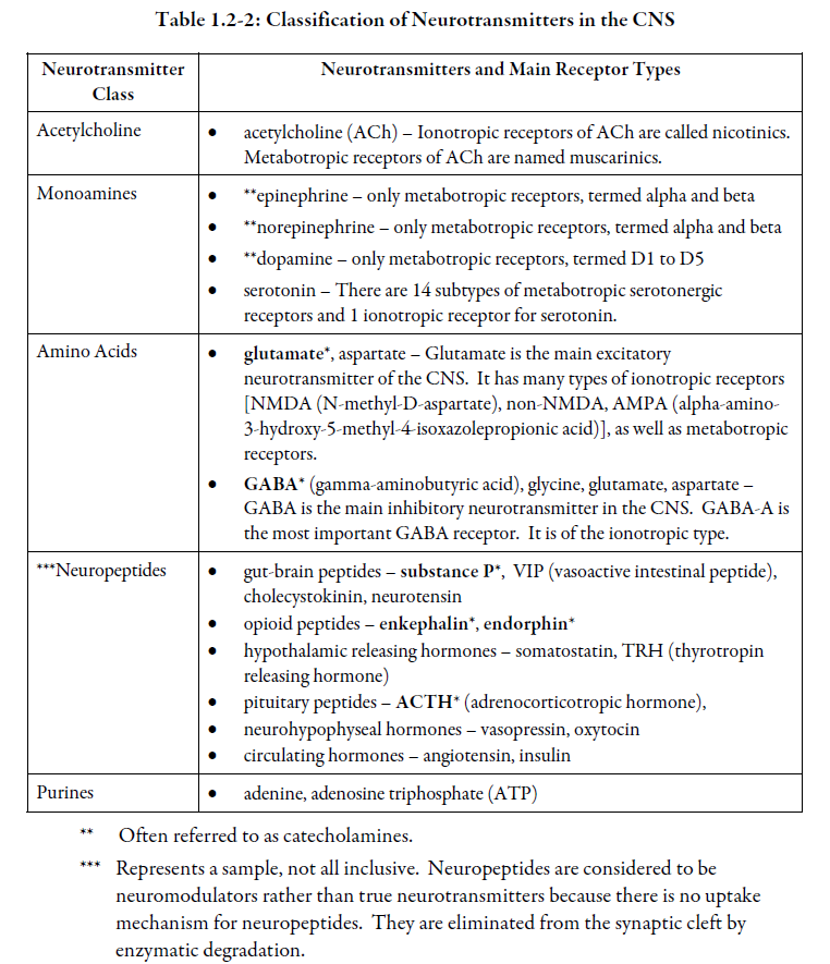
Classes of Neurotransmitters
Neurotransmitters are classified according to their chemical nature. Along with classical neurotransmitters, numerous molecules modulate neurotransmission but are not considered as true neurotransmitters. These substances are called neuromodulators.
Some neurotransmitters activate neurotransmission, and are referred to as excitatory neurotransmitters, while others reduce neurotransmission and are referred to as inhibitory neurotransmitters.
There are two classes of neurotransmitter receptors:
-
-
- ionotropic receptors*– These receptors form ion channels (ligand-gated ion channels). When neurotransmitters interact with this class of receptor, it is referred to as fast transmission because electrical properties of the cell are modulated right away.
- metabotropic receptors* – These receptors are coupled to second messengers that initiate intracellular signalling events that will modify cell metabolism. They are also referred to as G protein-coupled receptors (GPCR). When neurotransmitters interact with this class of receptor it is referred to as slow transmission because electrical properties of the cell are not modulated right away (modulation will occur ultimately, because most of these signalling events alter the state of activity of ion channels).
-
Each neurotransmitter possesses many types of receptors. However, not all receptor types are present in the same brain area or target organs. Specific receptor subtypes are discussed in more detail in pharmacological sections later in the course.
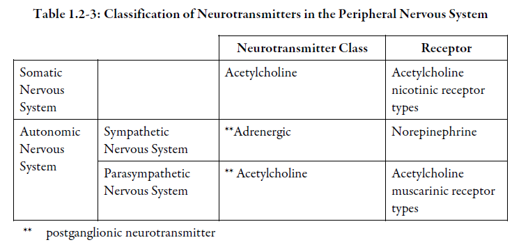
Some neurotransmitters act as both central and peripheral nervous system transmitters. In the somatic nervous system, acetylcholine is the neurotransmitter for all muscles that control body movement (somatic muscles). In the autonomic nervous system, the two different branches, sympathetic nervous system and parasympathetic nervous system, innervate most organs and produce opposing effects. The net effect of the two systems on a particular organ is referred to as the resting tone within that organ. See Tables 1.2-2 and 1.2-3.
Generally, each nerve cell releases only one type of neurotransmitter; however, some presynaptic neurons can release one or more neuromodulators. It is believed that differences in the frequency of nerve stimulation probably control which substance is released.
Did You Know?
As many as 100 or more different substances act as neurotransmitters or neuromodulators. However, only a little more than 20 have been well studied with respect to their actions.The German biologist Otto Loewi won the Nobel prize in 1921 for the discovery of the first substance acting as a neurotransmitter, acetylcholine.
Neurotransmitters produce their effects by interacting with receptors on the surface of the receiving cell. The shape and electrical charge of the transmitter and its receptor are felt to form the basis for the specificity of the transmitter-receptor interaction.
Each neurotransmitter tends to exert a characteristic action – excitatory or inhibitory, quick or slow, short or long lasting – but sometimes a neurotransmitter may behave differently at different sites. For example, acetylcholine exerts excitatory effects in the brain but in some portions of the peripheral nervous system, such as the heart, it has inhibitory effects.
As previously mentioned, once the neurotransmitter has exerted its effect, the action of the substance is terminated by degradation or re-uptake of the neurotransmitter. See Figure 1.2-4.
Although each neuron is generally assumed to release only one neurotransmitter, a single neuron may possess receptors for a variety of different neurotransmitters. This enables a neuron to receive impulses from many other neurons, each capable of exerting a different effect on the receiving neuron.
Cholinergic Neurotransmitters
One of the best known neurotransmitters is acetylcholine (ACh). Acetylcholine is synthesized from choline and acetyl coenzyme A by the enzyme choline acetyltransferase, and is broken down by enzymes acetylcholinesterase* and plasma cholinesterase*.
In the ANS, the cholinergic neurons include:
-
-
- all sympathetic and parasympathetic preganglionic neurons
- sympathetic postganglionic neurons that innervate most sweat glands
- all parasympathetic postganglionic fibers
-
Acetylcholine is the main transmitter for the parasympathetic system, and has important effects in the brain, including memory function.
As we mentioned previously, acetylcholine is the transmitter for all muscles that control body movement (somatic muscles). It is synthesized in spinal cord motor neurons, transported to peripheral nerve endings and released from vesicles to excite somatic muscles.
Acetylcholine has various roles as a transmitter in the periphery:
-
-
- excites activity – e.g., in somatic muscle and smooth muscle
- inhibits activity – e.g., slows the activity of pacemaker tissue in the heart which causes decreased heart rate (ANS)
- stores energy via digestion (ANS)
-
This diversity of activity points out the fact that each transmitter is not limited to a single behavioural identity, even though there is a clear association with a specified portion of the nervous system.
Adrenergic Neurotransmitters
The postganglionic sympathetic nervous system, in general, is described as adrenergic. The chief neurotransmitter at sympathetic neuroeffector junctions (released by postganglionic sympathetic fibres) is norepinephrine* (noradrenaline). Neurons that release norepinephrine are referred to as adrenergic.
Norepinephrine, epinephrine* and dopamine are classified chemically as catecholamines* and with serotonin*, form the monoamine neurotransmitter class.
1) Norepinephrine (NE)
Norepinephrine (NE) is derived from the amino acid tyrosine through a number of intermediary compounds. Its immediate precursor is dopamine. The action of norepinephrine is terminated by reuptake or by the enzymes catechol-O-methyl transferase (COMT)* and monoamine oxidase (MAO)*.
Like acetylcholine, NE is a neurotransmitter with an important role in the central and peripheral nervous systems to bring about action at effector organs.
2) Epinephrine
Epinephrine (or adrenaline*) is synthesized from NE. Contrary to NE, adrenaline is mostly found in the PNS. For example, in the sympathetic nervous system, adrenaline mobilizes the body against threatening, arousing or stressful events (“fight or flight”).
Did You Know?
There are multiple receptors for norepinephrine. The subtypes can be classified into alpha and beta-receptors, which are further subdivided into alpha-1, alpha-2, beta-1 and beta-2 receptors. Different subtypes of these receptors can be found on the presynaptic neuron as well as the postsynaptic neuron. The presynaptic alpha-2 receptor serves as an autoreceptor that will turn off further release of norepinephrine when norepinephrine binds to it.
Dopamine (DA)
In dopaminergic neurons, the amino acid tyrosine is converted by the enzyme tyrosine hydroxylase to L-DOPA* which is in turn converted to dopamine by a decarboxylating enzyme. Neurons using dopamine as a neurotransmitter are referred to as dopaminergic.
The action of dopamine is terminated by re-uptake or metabolism by the enzymes catechol-O-methyltransferase (COMT) or monoamine oxidase (MAO).
Dopamine is found in the brain and in nerves controlling peripheral tissues such as some blood vessels. In the CNS, DA is associated with various brain activities, such as control of body movements, mood, motivated behaviours and neuroendocrine functions (e.g., prolactin secretion is prohibited by dopamine). A deficiency of dopamine can cause disease characterized by motor dysfunction, e.g., Parkinson’s disease.
Did You Know?
Several types of dopamine receptors have been identified. They regulate dopamine neurotransmission. They may be found on the presynaptic neuron where they act as an autoreceptor or may be found postsynaptically. There are five subtypes of dopamine receptors: D1, D2, D3, D4 and D5. The most studied is the D2 receptor because it is stimulated by dopamine agonists for treatment of Parkinson’s disease and is inhibited by dopamine antagonists for treatment of schizophrenia.
Serotonin (5-Hydroxytryptamine or 5-HT)
Serotonin (5-HT) is synthesized from the amino acid tryptophan by the enzyme tryptophan hydroxylase. Neurons using serotonin as a neurotransmitter are referred to as serotonergic.
The action of serotonin is enzymatically terminated by monoamine oxidases. Reuptake is also an important mechanism to terminate 5-HT activity.
Serotonin has a number of effects in the body. It can be found in blood platelets*, the lining of the gastrointestinal tract, the brain, and smooth muscle of the cardiovascular system.
There are relatively few serotonin neurons in number, but they appear to be of major importance in the regulation of mood states, behaviour, thermoregulation, sleep, feeding, sexual behaviour and pain perception.
Amino Acid Neurotransmitters
1) Gamma-aminobutyric acid (GABA)
GABA is produced from the amino acid glutamate by the enzyme glutamic acid decarboxylase. Its action is terminated by re-uptake or by enzymatic degradation by GABA transaminase.
GABA controls the flow of neurochemical impulses by opening chloride channels that produce a hyperpolarization of the nerve membrane. Consequently, this inhibits or blocks nerve impulse propagation. GABA activity is increased by some drugs such as benzodiazepines and anticonvulsants.
GABA is the most common inhibitory neurotransmitter in the brain and overall may account for transmission in 25% to 40% of all brain synapses.
GABA plays an important role in various neurological and psychiatric disorders.
2) Glutamate
Glutamate is a major excitatory neurotransmitter in the CNS. Excessive activation of glutamatergic synapses causes large influxes of calcium into neurons, which can lead to cell death (referred as excitotoxicity).
This mechanism of cell death is believed to be important in acute neurological disorders such as stroke and CNS trauma.
Glutamate antagonists are being used therapeutically in the treatment of amyotrophic lateral sclerosis (ALS)* and dementia*.
Neuropeptide Neurotransmitters or Neuromodulators
Neuropeptides are proteins that consist of larger molecules than norepinephrine and acetylcholine, which are very small molecules.
Some neuropeptides are known to be both peripheral hormones and neuromodulators that act on the brain. For example, the neuropeptides enkephalins, endorphins and Substance P are involved in the perception of pain. Angiotensin II* induces a desire to drink when you lose body fluids via the renin-angiotensin-aldosterone pathway which controls sodium and water content in the body.
Did You Know?
A neuromodulator is a substance other than a neurotransmitter that is released by a neuron at a synapse, which can modulate cell excitability.A neurohormone is a hormone that is produced and secreted by neurons into the bloodstream, the cerebrospinal fluid, or the intercellular spaces of the nervous system.
A neuropeptide is composed of a short chain of amino acids (endorphins, enkephalins, vasopressin, etc.) found in brain tissue.
Purines
Adenosine triphosphate (ATP) is a purine nucleotide. It can be released together with ACh, dopamine and norepinephrine. Adenosine triphosphate has inhibitory action at intestinal smooth muscle and possibly in the brain.
Did You Know?
The bacteria Clostridium botulinum produces a toxin that blocks acetylcholine release at the neuromuscular junction. This causes muscle paralysis. A derivative of the botulinum toxin is used in aesthetic plastic surgery to produce skin muscle paralysis that reduces the appearance of wrinkles.
Summary — Section 2: Neurotransmission
Neurotransmission refers to the transmission of an impulse along the axon of a neuron to target cells or tissues. A stimulus is received by the dendrites. If a threshold of excitability is reached, an action potential is created and the impulse is conducted toward the cell body and along the axon. The impulse moves in one direction along the axon, depolarising the membrane as it is transmitted along the axon.
Electrochemical stimulation at the terminal buttons causes synaptic vesicles to release neurotransmitters into the synaptic cleft. Neurotransmitters released by vesicles located in the presynaptic neurons regulate the passage of the electrical impulse from one neuron to another neuron, or from a neuron to a muscle or effector organs.
The neurotransmitter reaches a postsynaptic receptor on the dendrite of a receiving cell and initiates either an excitatory or inhibitory process in that receptor. Excitatory actions will perpetuate the stimulus through the receiving neuron. Inhibitory actions will prevent the nervous impulse from moving through the next neuron.
After interacting with the receptor on the receiving cell, the neurotransmitter is either destroyed by enzymes or recaptured by the synaptic vesicles and stored for further use.
Presynaptic receptors (autoreceptors) on the terminal buttons of some neurons monitor the concentration of neurotransmitter within the synaptic cleft. Autoreceptors provide feedback, which controls the further synthesis, release and degradation of neurotransmitters.
In this section, we highlighted the following groups of neurotransmitters, each of which has specific roles, both within the nervous system and on various muscles and glandular tissues:
-
-
- cholinergic – e.g., acetylcholine
- adrenergic – e.g., norepinephrine, epinephrine
- dopaminergic – e.g., dopamine
- serotonergic – e.g., serotonin
- amino acids – e.g., GABA, glutamate
- neuropeptides – e.g., enkephalins, endorphins, substance P, angiotensin II
- purines – e.g., adenine, ATP
-
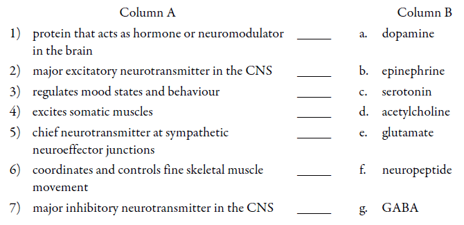
Progress check — section 2: Neurotransmission
1.
Indicate whether each of the following statements is True or False.
2.
Define action potential.
____________________________
____________________________
3.
Indicate whether each of the following statements is True or False.
4.
List 5 abnormalities that may occur with neurotransmitter systems.
1) ____________________________
2) ____________________________
3) ____________________________
4) ____________________________
5) ____________________________
5.
Match the description in Column A with the correct neurotransmitter in Column B.
6.
Which of the following neurotransmitters is inactivated by MAO? Select all that apply.
a. acetylcholine
b. dopamine
c. GABA
d. glutamate
e. norepinephrine
f. serotonin
7.
Which of the following neurotransmitters is classified chemically as a catecholamine? Select all that apply.
a. acetylcholine
b. dopamine
c. GABA
d. glutamate
e. norepinephrine
f. serotonin
Progress check answers — Section 2: Neurotransmission
1.
1) True
2) False
A synapse may be excitatory or inhibitory.
3) True
2.
An action potential is an electrical impulse that occurs when a neuron sends information down an axon, away from the cell body. An action potential is initiated by depolarization of the nerve cell.
3.
1) True
2) False
Each neuron usually releases only one type of neurotransmitter, but some presynaptic neurons can also release additional neuromodulator substances.
3) True
4) True
5) True
4.
1) not synthesized at all
2) synthesized in insufficient amounts
3) synthesized in excessive amounts
4) decreased numbers of receptors or receptor insensitivity
5) not degraded or taken back up into the presynaptic vesicles at the normal rate
5.
1) f. neuropeptide
2) e. glutamate
3) c. serotonin
4) d. acetylcholine
5) b. epinephrine
6) a. dopamine
7) g. GABA
6.
b. dopamine
e. norepinephrine
f. serotonin
7.
b. dopamine
e. norepinephrine
Test
1.
The autonomic nervous system is part of the ______________________ nervous system.
2.
The fight or flight response is promoted by the ______________________ branch of the autonomic nervous system.
3.
Which of the following statements regarding the axon of a neuron is true? Select all that apply.
a. has a special membrane that lets in some charged particles (ions) and keeps out others
b. is the cell body of the neuron
c. is not involved in the transmission of nerve impulses
d. may contain myelin on its outer surface
4.
An afferent or sensory nerve carries impulses:
a. owards the CNS
b. away from the CNS
5.
The junction between the dendrite of one neuron and the axon terminal of another is called a(n) ______________________.
6.
The electrical impulse that travels along a neuron is called a(n) _____________________.
7.
What cellular structure stores and releases neurotransmitters?
_____________________________
_____________________________
8.
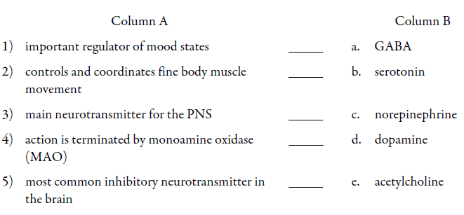
Match the description in Column A with the correct neurotransmitter in Column B. Neurotransmitters in Column B may be selected more than once. Descriptions in Column A may describe more than one neurotransmitter.
Test Answers
1.
peripheral
2.
sympathetic
3.
a. has a special membrane that lets in some charged particles (ions) and keeps out others
b. may contain myelin on its outer surface
4.
a. towards the CNS
5.
synapse
6.
an action potential
7.
Neurotransmitters are stored and released from vesicles located in the terminal axon of presynaptic neurons.
8.
1) b. serotonin
2) d. dopamine
3) e. acetylcholine
4)
b. serotonin
c. norepinephrine
d. dopamine
5) a. GABA
References
Section 1: Organization of the Nervous System
Vander, Sherman and Luciano. Human Physiology: the mechanism of body functions, Chapter 6, Structure and function of the nervous system, 10th Edition, McGraw-Hill, 2004.
Section 2: Neurotransmission
Goodman & Gilman. The Pharmacological Basis of Therapeutics, Chapter 6, Neurotransmission, 11th Edition, McGraw-Hill, 2006.
Mathews GG. NEUROBIOLOGY Molecules, Cells and Systems. Available at URL http://www.blackwellpublishing.com/matthews/animate.html
Animations showing channel gating during an action potential, propagation of the action potential, synaptic vesicle fusion and neurotransmitter release, and comparison of direct and indirect neurotransmitter actions.
Vander, Sherman and Luciano. Human Physiology: the mechanism of body functions, Chapter 6, Structure and function of the nervous system, 10th Edition, McGraw-Hill, 2004.